

Warm welcome, fellow internet user, to this highly specialized space for general surgery, hepatobiliary pathology, diagnostic ultrasound, and preventive health.
Interpreting the finding of a hepatized gallbladder in an ultrasound report requires going beyond routine reading and understanding how the accumulation of dense content modifies the acoustic impedance of the gallbladder cavity.
As a specialist surgeon and tenured professor at the University of Cuenca, my priority is to provide you with absolute peace of mind and medical precision, teaching that this ultrasound pattern is observed when bile is replaced by hyperdense biliary sludge, pus, or bloody remains that simulate liver tissue.
Before deconstructing the difference between a benign swollen mass and a primary gallbladder tumor, analyzing why changes in patient position during ultrasound clarify the diagnosis, or explaining how our cutting-edge protocol addresses high-difficulty cholecystectomy, I invite you to seek certainty in the scientific evidence.
Under the academic direction of Dr. Jorge Delgado, we evaluate your hepatobiliary health with the utmost clinical rigor. This time, without further ado, let's delve into the topic of study proposed in a forum on a chronic disease: hepatized gallbladder.
To understand this, we will first define the gallbladder. This is a small, pear-shaped organ that plays an essential role in digestion, as it stores, concentrates, and releases bile (30 to 50 ml in 24 hours), a greenish-yellow fluid produced in the liver. It helps break down fats in the small intestine.
However, rare chronic conditions, such as hepatized gallbladder, can cause serious complications and discomfort in the biliary system.
Common doubts
What exactly does it mean to find a hepatized gallbladder on ultrasound?
It is an ultrasound phenomenon where the interior of the gallbladder loses its anechoic (black) content and shows a homogeneous or heterogeneous echogenicity similar to that of the adjacent liver tissue, giving the false visual impression that the gallbladder has "turned into a liver".
What are the main causes that produce this pattern of hepatization?
Tumefactive biliary sludge: severe accumulation of cholesterol crystals and hyperviscous bile (most common benign cause).
Pyocholecystitis / Empyematous cholecystitis: massive accumulation of pus and bacterial debris due to severe infection.
Hemocholecyst: clotted blood within the gallbladder lumen secondary to trauma, biopsy, or hemorrhage.
Gallbladder cancer: infiltrating gallbladder adenocarcinoma that occupies and destroys the entire cavity.
Why does this finding represent a risk of false diagnosis in ultrasound?
Because an inexperienced operator may mistake the hepatized gallbladder for a normal extension of the liver parenchyma or for a lobe of the liver, mistakenly reporting that "the gallbladder is not visualized due to previous cholecystectomy" or that "the gallbladder is sclerotic," overlooking a serious infection or an active tumor.
What additional tests clarify whether the cause is biliary sludge or a tumor?
Color Doppler ultrasound: if the internal mass does not show vascular blood flow, it is likely biliary sludge or pus. If it shows internal vascularization, neoplasia is suspected.
Decubitus Maneuver (Mobility): observe if the contents shift when moving the patient from position.
Computed Tomography (CT) or Magnetic Resonance Imaging (MRI): Essential when there is doubt about parietal infiltration or malignancy.
If you came to this blog because of abdominal pain or simply to obtain new specialized information for your personal development, which allows you to maintain a proactive attitude towards health problems.
This proactive determination ensures that you seek medical attention at hospitals or health clinics within the public or private healthcare system, according to your personal geographical location, in order to reach an early diagnosis and avoid serious complications from self-assessment and, even worse, from self-medication; all they do is delay the assistance of medical personnel in your locality.
In this article you will find everything you need to know through suggestions " written by people, for people" and, at the same time, we will talk about the causes, symptoms and treatment options of hepatized gallbladder, as well as how to treat it through advanced medical or surgical procedures.
Hepatized Gallbladder
Hepatized gallbladder is a relatively uncommon phenomenon; it is usually due to an underlying chronic condition, such as cholecystitis (inflammation of the gallbladder) or pancreatitis (irritation of the pancreas), although it can also arise from an obstruction of the biliary tract, which determines the chronic presence of bile of higher concentration, as well as the formation of biliary sludge or mud stuck to the wall of the gallbladder.
Symptoms are usually intense and may include nausea and vomiting, abdominal pain and tenderness, fever, or chills.
Complications arising from a hepatized gallbladder can be serious; affected individuals may suffer gallbladder perforation, with chemical peritonitis and, in some cases, death from sepsis (contamination of the blood throughout the system) if not treated promptly.
Fortunately, treatment usually consists of broad-spectrum antibiotics to control and reduce inflammation and infection of the abdominal cavity and bile ducts; those with severe cases require surgery to remove the gallbladder and complete the treatment.
Therefore, although hepatized gallbladder is a rare condition with potentially serious consequences, medical checkups that allow for early and timely diagnosis and treatment can help minimize the risk of serious complications.
Causes of hepatized gallbladder
Hepatized gallbladder is a chronic condition that occurs when the liver produces excess bile due to poor diet (too much fat, fried foods and sodas), leading to the formation of "biliary sludge . "
In the ultrasound examination of the gallbladder, it is this heavy bile that presents ultrasound characteristics similar to those of the hepatic gland and the hepatized gallbladder.
In most diseases of the biliary viscera, there is an ultrasound difference between the liver tissue, the gallbladder wall and the gallstones (lithiasis), which is an important fact to consider in the general analysis of the complementary examinations.
Furthermore, chronic degenerative diseases originating from liver inflammation and the accumulation of scar tissue in the liver, as well as in the walls of the gallbladder, are characterized by marked liver dysfunction. There are several causes of hepatized gallbladder scarring, including:

- Chronic liver disease: People with chronic liver disease, such as cirrhosis, have a higher risk of developing hepatized gallbladder.
- Alcohol abuse: Excessive alcohol consumption damages the structure of the liver and contributes to the development of the biliary tract.
- Poor diet and nutrition: A diet high in saturated and trans fats, artificial colors and flavorings (petroleum derivatives) causes permanent inflammation of the liver with increased bile production for detoxification; therefore, more heavy bile (biliary sludge or mud) accumulates on the walls of the gallbladder, which makes it difficult for the gallbladder to contract.
- Infections: Viral infections, such as hepatitis B and hepatitis C, can cause chronic fibrosis in the liver and, consequently, in the biliary tract.
Symptom
A hepatized gallbladder can cause a range of symptoms that affect daily quality of life. Some of the most common manifestations of a hepatized gallbladder are:
- persistent pain: It is usually felt in the upper right quadrant of the abdomen, but it can also radiate to the back, at the level of the base of the right shoulder blade.
- Nausea and vomiting: Patients with hepatized biliary viscera may experience episodes of nausea and vomiting, which can lead to dehydration and weight loss.
- Bloating and gas: A hepatized gallbladder can cause bloating due to the accumulation of gas in the stomach and intestines because of poor fat digestion.
- Loss of appetite: Patients may lose their appetite or feel full after eating small portions.

Diagnosis of Hepatized Gallbladder
Hepatic or hepatized gallbladder is a rare but serious condition that can be difficult to diagnose.
It occurs when the gallbladder becomes inflamed and thickened due to prolonged obstruction of the cystic duct or the common bile duct ; in turn, this causes a chronic accumulation of bile, giving it a "hepatized" appearance.
Diagnosis typically requires imaging studies performed by an experienced radiologist, as well as laboratory results interpreted by a pathologist. Hepatized gallbladder often presents with symptoms similar to those of cholecystitis, such as abdominal pain, nausea, vomiting, and fever.
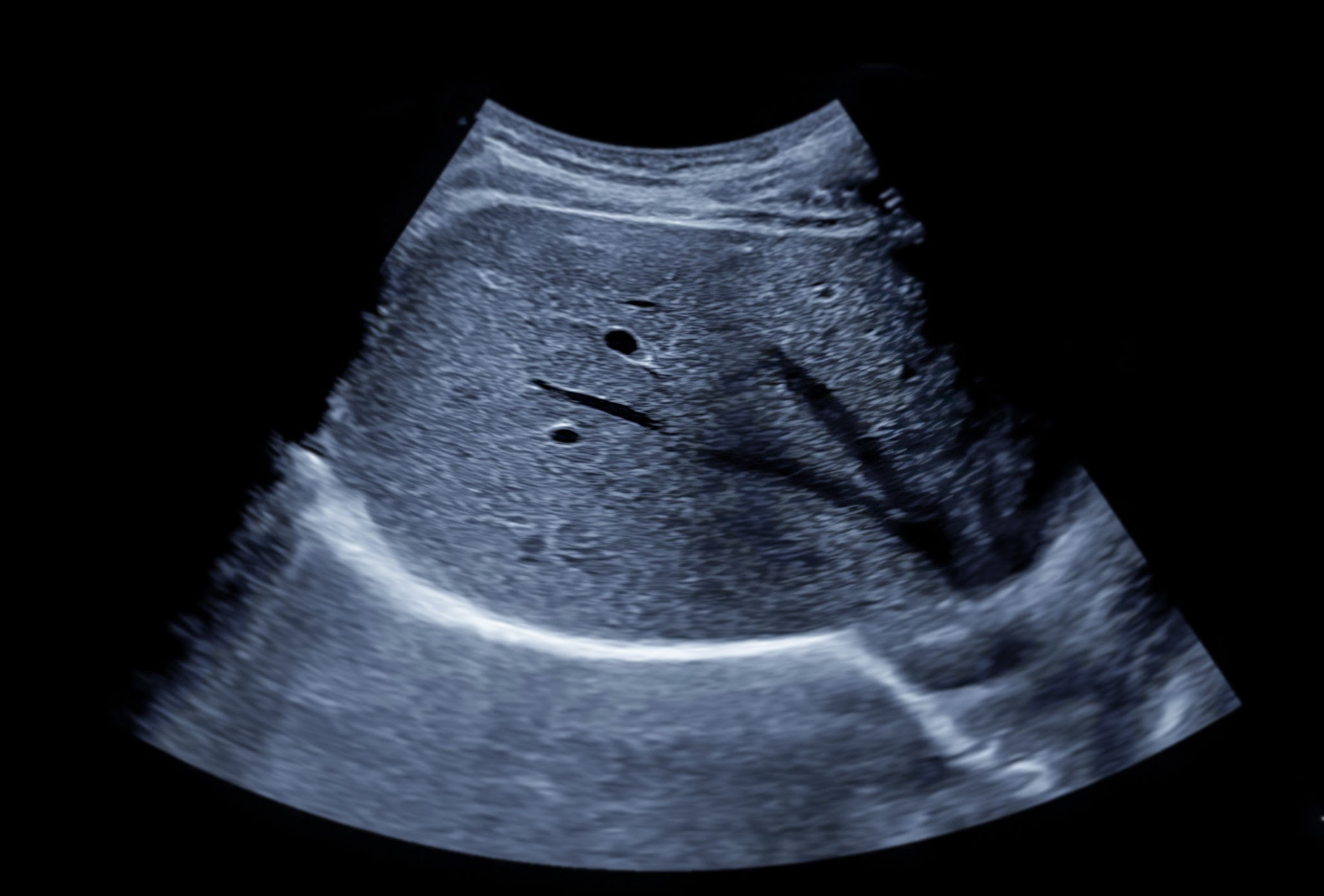
The most reliable way to diagnose this condition is endoscopic retrograde cholangiopancreatography (ERCP), which allows visualization and simultaneous treatment of the underlying cause of the obstruction.
If not treated promptly, hepatized gallbladder can lead to serious complications, such as sepsis or even death; therefore, prompt diagnosis and appropriate medical intervention are essential.
⚠️ ATTENTION:
This content is for informational purposes only. For medical advice or diagnosis, consult a healthcare professional in your area.
Differential diagnosis
Hepatized gallbladder, also known as the vesicular variant of hepatization, is a rare condition caused by an abnormal inflammatory process that usually occurs in people with chronic gallbladder infection that is not treated promptly.
Although this condition can be difficult to diagnose correctly, there are certain key factors and characteristics that medical professionals use to differentiate hepatized gallbladder from other types of gastrointestinal infections.
These usually include ultrasound findings such as thickening of the walls , changes in the shape and size of the gallbladder, and other associated symptoms, such as pain in the right upper quadrant, fever, nausea and vomiting, jaundice, etc.
Physicians may also choose to perform laboratory tests to confirm that hepatized gallbladder is the underlying cause of the patient's illness. Differential diagnosis remains an important aspect of quickly and accurately identifying and treating this rare infection.
Another distinguishing characteristic is intense pain in the upper right region of the abdomen. Sometimes, this corresponds to a functional disorder of the gallbladder wall, with marked dilation of the gallbladder.
It is a characteristic of the thin-walled acalculous gallbladder (without stones inside), with a similar variety of discomforts.
Treatment options
Treatment depends on the severity of the condition and its underlying causes . Here are some ways to manage and treat hepatized gallbladder:
- Changes in lifestyle: Make changes to your diet and lifestyle, such as reducing alcohol consumption and increasing your intake of dietary fiber, fruits, vegetables and fresh greens, They help control the production of heavy bile that is deposited on the walls of the gallbladder, and it should also be remembered that this heavy bile is gallstone-forming.
- Medication: Your trusted local doctor can prescribe medication to control digestive symptoms such as nausea and vomiting.
- Surgery: In severe cases, surgical intervention may be necessary, preferably minimally invasive laparoscopic surgery, to remove the gallbladder and treat hyperlipidemia (excess cholesterol, polyunsaturated and trans fats, and a lack of fat-soluble vitamins).
In this section of the article, we will dedicate ourselves to answering questions posed by patients who come to the doctor's office or to the emergency room:
To know if I have liver or gallbladder problems
When determining whether you have a liver or gallbladder disease, there are certain symptoms and signs that can help diagnose it. Hepatized gallbladder is a condition in which chronic inflammation, infection, or obstruction causes the gallbladder to fill with bile from the liver.
Symptoms may include pain in the upper right side of the abdomen, nausea and vomiting, fever or chills, jaundice (yellowing of the skin and eyes), and clay-colored stools.
If any of these symptoms occur, it is important to see a doctor immediately, as a hepatized gallbladder can lead to serious complications if left untreated.
A healthcare professional in the public or private system will assess your symptoms and perform tests, such as imaging scans or blood tests, to determine if you have a hepatized gallbladder or another type of liver or biliary problem.
Based on what they find when they take a complete medical history followed by a thorough physical examination of the abdomen and analyze the additional tests, they may recommend lifestyle changes, medications, or surgery to treat your condition.
Symptoms present when the gallbladder is damaged
Hepatized gallbladder is a condition caused by damage or disease of the gallbladder, which causes inflammation due to the arrival of heavy bile from the liver and in the gallbladder due to its ability to concentrate bile. We find extra heavy bile that sticks to the wall of the gallbladder and gives it the appearance of liver tissue when performing an ultrasound, as well as alteration in the contraction of the same.
Although it can be a serious problem, it often goes unnoticed for some time, as there are few common symptoms associated with it, such as a slight discomfort, followed by indigestion, after consuming fatty foods.
Over time, these symptoms may include abdominal pain in the upper right side, nausea, vomiting, jaundice (yellowing of the skin), fever, and chills. If left untreated, a hepatized gallbladder can lead to serious complications, such as sepsis or even death.
Therefore, it is essential that people experiencing any of its associated symptoms seek medical advice so that diagnostic tests can be ordered and appropriate treatments administered.
Prevention of hepatized gallbladder
The hepatized gallbladders are an important component of the pathophysiology of the liver and biliary tract. Unfortunately, when these gallbladders are damaged due to chronic inflammation from excessive bile, gallstones, or other conditions, they can lead to serious health problems, such as fatty liver disease and hepatitis.
The good news is that prevention is possible, particularly through measures to reduce obesity, sedentary lifestyles, and other unhealthy habits. Physical exercise, such as walking, jogging, swimming, and cycling, helps keep the body moving.

Following a balanced diet with plenty of dietary fiber from fruits and vegetables, exercising regularly to strengthen muscles, and staying active through a fitness program are excellent ways to prevent hepatized gallbladders from becoming damaged over time.
Proactive measures like these are key to being the protagonist that helps keep your liver healthy for years and, at the same time, is reflected in good overall health.
What are the radiological features on contrast-enhanced computed tomography (CT) that differentiate swollen biliary sludge from gallbladder adenocarcinoma?
Differentiating between tumefactive biliary sludge and gallbladder adenocarcinoma using triphasic computed tomography (CT) with intravenous contrast represents one of the finest challenges in hepatobiliary diagnosis.
The absolute key lies in the vascular behavior : biliary sludge is an avascular mass that does not enhance with contrast, while the neoplasm presents vascular neoformation and contrast retention due to its tumor stroma.
| Radiological Criteria (CT scan) | Swollen Biliary Sludge (Benign) | Vesicular Adenocarcinoma (Malignant) |
| Enhancement with Contrast IV | Absent (0 UH increase). No sign of vascularization at any stage. | Heterogeneous and Intense. Peripheral/nodular enhancement in the arterial phase and retention in the late phase. |
| Attenuation Without Contrast | Homogeneous or slightly hyperdense (20–50 HU), higher than normal bile. | Iso to hypodense relative to the hepatic parenchyma (30–40 HU), with areas of necrosis. |
| Adjacent Vesicular Wall | Thin, uniform (< 3 mm) and intact. Maintains wall stratification. | Thickened asymmetrically and irregularly (> 5 mm), with loss of stratification. |
| Interface with the liver | Subserous fat plane (perivesicular plane) preserved and clear. | Invasion of the hepatic bed. Loss of the cut plane corresponding to segments IVb and V of the liver. |
| Gravity Effect / Level | It adapts to the decline or forms a fluid-liquid level after resting. | Fixed, rigid mass, not dependent on the patient's position. |
| Vascular and Lymph Node Involvement | Absent. No lymphadenopathy or hilar involvement. | Lymphadenopathy in the hepatoduodenal ligament and possible involvement of the portal vein and/or hepatic artery. |
Key Points of Tomographic Interpretation
-
Hounsfield Unit (HU) measurement: The placement of the Region of Interest (ROI ) before and after contrast administration is the key step. An increase of less than 10 HU confirms the vascular nature of the swollen biliary sludge. An increase of 20 HU is indicative of solid neoplastic tissue.
-
The late stroma sign: gallbladder adenocarcinoma contains abundant desmoplastic fibrous tissue. In the late or washout phase (3 to 5 minutes post-injection) , the contrast agent washes out of the liver but remains "trapped" within the gallbladder tumor mass, enhancing its contrast against the adjacent hepatic parenchyma.
-
Biliary Tract Invasion: The presence of isolated dilation of the intrahepatic bile ducts (neoplastic Mirizzi syndrome) adjacent to a gallbladder mass confirms the infiltrative nature of the carcinoma.
Value analysis
The Semiological Maneuver of Mobility and Compression by Transducer:
Dr. Delgado brings a unique value of high-level teaching by teaching the swollen mud test in real time.
Teaching that by gently pressing with the ultrasound transducer or by placing the patient in the left lateral decubitus position, dense biliary sludge shows a subtle "deformation or slow laminar flow" without a Doppler signal, while a tumor is rigid, does not deform and captures a vascular signal, gives the article an unsurpassed semiological mastery.
Academic and Professorial Vision (Department of Surgery – University of Cuenca):
The differential diagnosis is reinforced when faced with the surgical challenge of a hepatized pyocholecyst . As university professors, we teach residents that when operating on a hepatized gallbladder, the wall is usually friable and Calot's triangle obliterated.
The prior endoluminal puncture and irrigation to evacuate the hyperdense contents constitute the indispensable anatomical step to achieve adequate traction and expose the Strasberg Safety Critical View without injuring the main bile duct.
Conclusion
Finally, it's important to note that hepatitis is a condition that can cause discomfort and affect daily life. It's essential to monitor the symptoms, as they are " written by people, for people ," and to seek medical attention if a gallbladder condition is suspected.
Treatment for gallbladder hepatization requires a combination of lifestyle changes, medication, and, in severe cases, surgery. If you experience any symptoms of gallbladder hepatization , talk to your doctor for proper diagnosis and treatment.
Now is the time to share preventative information on social media so that more people in your circle can benefit from this news and help reduce the serious complications of a diseased gallbladder.
Thank you for leaving your comment and suggestions for the blog's audience on digestive health. Warm regards, and see you in the next post!
Dr. Jorge Delgado, Surgeon. Hepatized Gallbladder.
📌 Recommended readings:
🎯 Direct contrast with normal gallbladder anatomy and wall integrity.
🎯 Analysis of hydrops, mucocele and intraluminal hyperpressure.
🎯 Lesson on perforation, necrosis and subhepatic phlegmon.