

Hello, dear netizen, and welcome to this highly specialized space in general surgery, hepatobiliary reconstruction, diagnostic ultrasound and preventive health.
Facing the ultrasound finding of a gallbladder full of stones (calculi) requires overcoming visual shock and understanding the severe functional implications of massive cholelithiasis.
As a specialist surgeon and full professor at the University of Cuenca, our priority is to teach you that a gallbladder full of stones has lost its normal contractile dynamics and acts as a focus of progressive chronic inflammation.
Before deconstructing why microcalculi are the main cause of acute biliary pancreatitis, analyzing the risks of gallbladder sclerosis, or explaining how our cutting-edge protocol addresses high-difficulty laparoscopic cholecystectomy using Strasberg's Critical Safety View, I invite you to seek certainty in the scientific evidence.
Under the academic direction of Dr. Jorge Delgado, we evaluate your hepatobiliary health with the utmost clinical rigor in preventive health, with a focus on diseases of the digestive system.
Common questions
What does it really mean to have a gallbladder full of stones on an ultrasound?
This means that the gallbladder cavity is largely or entirely occupied by cholesterol, pigment, or mixed gallstones.
This prevents the gallbladder from filling and emptying properly to digest fats, turning it into a permanently inflamed and non-functional organ.
Why is a gallbladder full of small stones more dangerous than one with a single large stone?
Because small stones (microcalculi < 5 mm) can easily slide through the cystic duct into the common bile duct.
As they migrate, they block the drainage of the pancreas and bile, triggering acute biliary pancreatitis (a life-threatening complication) or choledocholithiasis with jaundice and infection (cholangitis).
Can gallstones be eliminated or dissolved with juices, oils, or medications?
No. It's an extremely dangerous myth. So-called "gallbladder washes or home remedies to expel stones" cause strong gallbladder contractions that force the stones into the main bile duct, triggering serious blockages in the ampulla of Vater and requiring emergency surgery.
What is the definitive treatment for a gallbladder full of gallstones?
The only curative treatment is laparoscopic cholecystectomy (surgical removal of the gallbladder).
By removing the diseased reservoir through millimeter incisions, the flow of bile from the liver to the intestine is directly restored, forever eliminating the risk of colic, pancreatitis, and gallbladder cancer.
Let's get started with the topic: a gallbladder full of stones, or biliary lithiasis. It's a condition that occurs when there are gallstones or stones in the gallbladder.
This condition occurs due to a chemical imbalance of the bile-forming components, thickening of the bile wall, and increased bile flow.
It can be caused by various factors, such as diseases like diabetes, cirrhosis, and Crohn's disease. Its symptoms are "written by people, for people" and can include sudden pain in the upper right abdomen, gas, nausea, vomiting, diarrhea, bloating, and a feeling of fullness in the stomach.
If you came to the preventive health blog because of discomfort or for new information for your personal development, this will help you maintain a proactive attitude towards abdominal pain and seek medical attention in hospitals or clinics, whether from the public or private health system, depending on your geographical location, and reduce complications.
A proactive attitude also confronts self-reporting and self-medication, two negative actions that delay a visit to the doctor and, as the hours or longer pass, complications are not long in coming.
It's time for a cup of green tea, a 100% natural antioxidant containing the amino acid L-theanine, which has a special property of improving concentration and information retention in these times of countless mental distractions. Let's delve into this refreshing read about gallstones!
Gallbladder full of stones
Gallstones are a surprisingly common disorder, with the Centers for Disease Control and Prevention estimating that up to 10% of Americans have them . This staggering prevalence underscores the importance of understanding their causes, symptoms, and potential complications.
Gallstones form when the gallbladder concentrates bile, causing excess cholesterol or bilirubin to crystallize inside. They can vary in size from tiny grains of sand to stones the size of a golf ball.
These gallstones can remain asymptomatic for long periods of time , but when they obstruct the ducts from the gallbladder (cystic) and the common bile duct to the intestine and cause a buildup of bile, they can lead to a painful attack called acute or chronic cholecystitis.
Although some people with gallstones have no symptoms, others may experience upper abdominal pain, nausea and vomiting, or jaundice. If left untreated, gallstones can lead to serious complications , such as inflammation of the pancreas, infection, and rupture of the gallbladder.
The only way to remove gallstones is through surgery, either by laparoscopic cholecystectomy (removal of the gallbladder) or by combining it with endoscopic retrograde cholangiopancreatography (ERCP) when the stones have passed into the common bile duct. Being aware of these potential risks is crucial for seeking preventive medical attention if you suspect you have gallstones.
Taking preventative health measures, such as eating healthy foods and exercising regularly, can help reduce the likelihood of developing gallstones. Knowing the facts about this condition can help you recognize its symptoms and seek prompt medical attention if needed.
Endoscopic retrograde cholangiopancreatography (ERCP)
Endoscopic retrograde cholangiopancreatography (ERCP) is a diagnostic and therapeutic procedure that combines endoscopy and radiology to visualize the bile ducts and pancreas.
Through an endoscope, a flexible tube equipped with a mini-camera and a light source, the duodenum, which is the beginning of the small intestine, is accessed, where the duodenal papilla (in the second portion of the duodenum), the opening of the bile and pancreatic ducts, is located.
Once the endoscope is inserted, a contrast medium is injected into the bile and pancreatic ducts using a specialized catheter. This allows for radiographic imaging via fluoroscopy, facilitating the detection of blockages, stones, strictures, tumors, or other abnormalities in the bile ducts and pancreas.
ERCP is particularly valuable for identifying and treating conditions such as cholelithiasis, pancreatitis, and tumors of the pancreas and bile ducts.
In addition to its diagnostic function, ERCP also allows for therapeutic interventions, such as the removal of gallstones, the placement of stents to relieve obstructions, or the biopsy of suspicious lesions.
Although generally safe, the procedure carries certain risks, including post-procedure pancreatitis, infections, bleeding, and perforations.
Therefore, ERCP should be performed by an experienced endoscopist and with adequate patient preparation, including evaluation of medical history and identification of contraindications.
ERCP is considered a key procedure in gastroenterology, as it offers both diagnostic and treatment options for complex conditions of the biliary and pancreatic systems.
⚠️ IMPORTANT NOTE:
This content is for informational purposes only. For medical advice or diagnosis, consult a healthcare professional in your area.
Causes of gallstones:
Firstly, bile is formed in the liver and its density depends on the type of diet provided by the individual; thus, a diet rich in fats and fried foods and poor in liquids, with the habit of a sedentary lifestyle, causes the liver gland to produce heavy bile.
Therefore, the presence of gallstones in the gallbladder is due to a chemical imbalance in the composition of bile. The function of the gallbladder is to further concentrate the bile coming from the liver.
This is how the excessive formation of gallstones begins, and it is also due to obesity and the modern diet, characterized by industrialized dry foods rich in artificial colors and flavorings.
In addition, excessive consumption of refined sugar and carbohydrates through pasta with artificial flavors and colors, alcohol, and coffee can precipitate and initiate the development of gallstones.
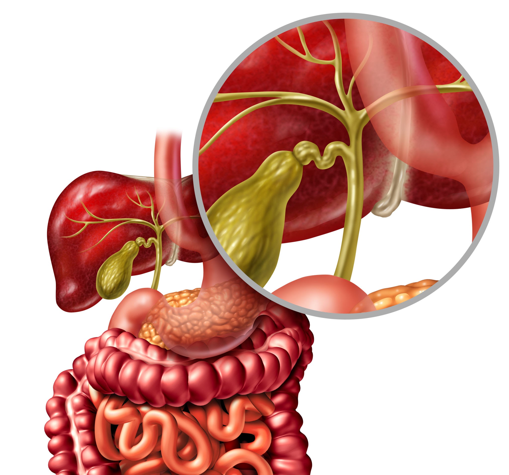
In addition to these factors, gallstone formation can also occur due to chronic diseases such as diabetes, cirrhosis, and autoimmune conditions such as Crohn's disease or irritable bowel syndrome, as well as acute inflammation of the gallbladder, called cholecystitis.
Symptom
The main discomforts associated with gallstones or biliary tract stones are sudden pain in the upper right abdomen, followed by nausea, vomiting and diarrhea, fever and loss of appetite; feeling of fullness in the stomach; abdominal bloating and gas; pains in the back and right shoulder; and greenish vomiting due to the high presence of bile.

Treatments
There are several treatment options for gallstones, and they will depend on the number and size of the stones in the gallbladder.
The main purpose of the treatment is to remove gallstones or stones in the biliary viscera by means of the technique known as cholecystectomy to avoid cholecystitis or inflammation of the gallbladder tissue.
When the stones or calculi are small, they can be eliminated through drug treatment.
These treatments include medications that relax the gallbladder to dilute the stones and facilitate their expulsion, or that increase bile flow to expel them.
In some cases where medical treatment is not enough, laparoscopic surgery to remove the gallbladder may be necessary when the stones or gallstones are larger and cannot be treated with drugs.
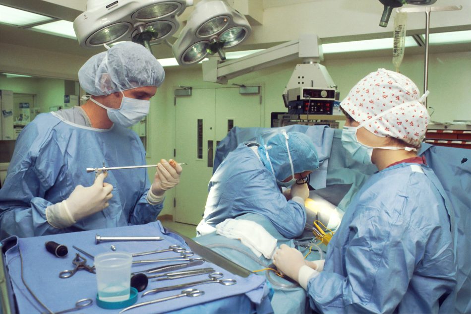
General anesthesia will be administered for the minimally invasive procedure to remove the gallbladder filled with gallstones . This laparoscopic surgery involves minimal incisions of 0.5 to 1 cm in length, allowing for a faster recovery than conventional or open surgery.
Differential diagnosis
The differential diagnosis of a gallbladder filled with stones includes several conditions that can present similar symptoms, such as:
Acute cholecystitis: Inflammation of the gallbladder, usually caused by obstruction by a gallstone.
Pancreatitis: Inflammation of the pancreas, which can be caused by gallstones that obstruct the pancreatic duct.
Peptic ulcer: injury to the lining of the stomach or duodenum that can cause similar abdominal pain.
Gastritis: Inflammation of the stomach lining that can cause pain and discomfort.
Hepatitis: Inflammation of the liver that can cause gastrointestinal symptoms.
Kidney stones: They can cause abdominal or back pain that is sometimes mistaken for biliary pain.
Irritable bowel syndrome: gastrointestinal disorder that can cause abdominal pain and changes in bowel habits.
Appendicitis: Inflammation of the appendix in a retrocecal position that can cause pain in the lower right part of the abdomen and be confused with biliary pain.
A complete clinical evaluation and imaging studies are essential to establish an accurate diagnosis and differentiate between these conditions.
Prevention
To prevent gallstone formation , it is recommended to follow a healthy , low - fat diet. This helps prevent bile from becoming thick, which can contribute to the formation of gallstones.
It is also recommended to avoid excessive consumption of sugar and carbohydrates, nuts, alcohol and coffee, as these have been shown to be contributing factors to gallstones.
In addition, maintaining a healthy weight is recommended to avoid obesity, which has also been identified as a risk factor for this disorder.
From a surgical and hepatobiliary perspective, massive cholelithiasis (gallbladder filled with multiple stones or "full of stones") represents a physiological time bomb.
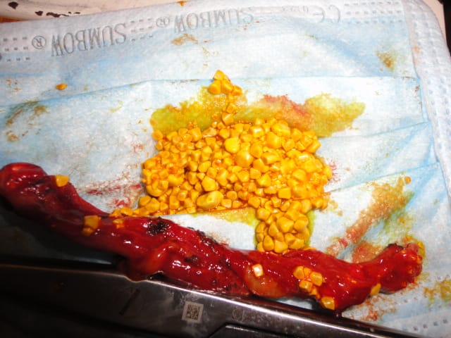
When the gallbladder reservoir is completely filled with microcalculi or faceted stones of various sizes, the gallbladder loses its ability to retract and suffers a permanent (chronic) mural inflammation process.
The real clinical danger lies not only in the pain of biliary colic, but also in the migration of microcalculi to the common bile duct (acute pancreatitis or choledocholithiasis) , extrinsic compression of the biliary tract ( Mirizzi syndrome ) or the evolution towards a scleroatrophic porcelain-walled gallbladder, with a high risk of neoplastic degeneration.
Mirizzi syndrome
Mirizzi syndrome is a severe and rare complication of long-standing cholelithiasis (present in 0,5% to 2,5% of patients operated on for biliary lithiasis).
It occurs when a large stone (or multiple stones) rigidly impacts the neck of the gallbladder or the cystic duct , triggering a chronic inflammatory response that produces extrinsic compression, ischemia, necrosis and even progressive erosion towards the common hepatic duct (CHC) or choledochus , generating a cholecystocholedochal fistula.
Csendes Surgical Classification (The Gold Standard)
To plan the operating room strategy and avoid the dreaded iatrogenic injury of the main bile duct, we follow the classification of Csendes et al. (modified with Type V by Beltrán):
| Mirizzi type | Anatomy and Degree of Injury | Recommended Surgical Management and Behavior |
| Type I | Extrinsic compression of the CHC due to impact on the cystic duct/mouth. Without fistula (biliary tract wall intact). | Cholecystectomy (total or subtotal) + exploration of the biliary tract or preventive placement of a Kehr tube if the wall is very friable. |
| Type II | Cholecystocholedochal fistula what affects less than 33 % (<1/3) of the circumference of the main bile duct. | Subtotal cholecystectomy (leaving a flap of the gallbladder wall to repair the fistula) + primary closure over the Kehr tube (T-tube). |
| Type III | Cholecystocholedochal fistula which affects between the 33% and 63% (1 / 3 the 2 / 3) of the circumference of the duct. | Subtotal cholecystectomy + complex repair with flap over the Kehr tube o biliodigestive bypass (hepatojejunostomy in Roux-en-Y). |
| Type IV | Complete destruction (> 66% o 100 %) of the wall of the main bile duct (the anterior and posterior walls are eroded). | Roux-en-Y hepaticojejunostomy (Biliary-digestive bypass is mandatory; primary repair over a Kehr is contraindicated due to narrowing). |
| Type V | Any type (I to IV) associated with a cholecystoenteric fistula (Va: without gallstone ileus; Vb: complicated by gallstone ileus due to intestinal migration of the stone). | Treatment of bilio-biliary fistula according to grade I-IV, PLUS Closure of the enteric defect (duodenum/colon) and enterolithotomy if there is obstruction. |
Tactical Principles of Rescue in the Operating Room
-
SOS – Avoid Blind Dissection of Calot's Triangle: In Mirizzi syndrome, the hepatocystic triangle is usually completely obliterated by a dense block of inflammation and fibrosis. Forcing traction to visualize the structures can lead to inadvertent transection of the common bile duct or hepatic artery.
-
Gallbladder Opening ( Fundus-First / Antegrade Technique): When a Mirizzi syndrome is suspected or found, the strategy of choice is incision of the gallbladder fundus (cholecystotomy) , evacuation of the stones from the inside and exploration of the anatomy from the inside out ( inside-out approach ).
-
Maintenance Subtotal Cholecystectomy: The entire posterior wall of the gallbladder should not be removed when it is adhered to the liver bed or the biliary tract. Leaving the posterior wall attached and electrocauterizing the mucosa preserves the structural integrity of the biliary tract and provides the autologous tissue necessary for tension-free reconstruction.
Value analysis
The Risk of Sclerotic Gallbladder and Mirizzi Syndrome:
Dr. Delgado brings a unique value of high surgical training when addressing the technical challenge of long-standing cholelithiasis.
Teach that a gallbladder "full of stones" that is not operated on in time suffers progressive fibrosis, contracting rigidly around the stones ( scleroatrophic gallbladder ) and inducing inflammation of the hepatoduodenal ligament.
This can cause erosion of a large stone into the common bile duct ( Mirizzi syndrome types I-IV ), turning a simple cholecystectomy into a complex biliary reconstruction.
Academic and Professorial Vision (Department of Surgery – University of Cuenca):
The difference is reinforced by Strasberg's Critical Vision Safety (CVS) teaching . As a university professor, I teach residents and interns that, when faced with an inflamed, thickened, or sclerotic gallbladder, the anatomical key to preventing iatrogenic biliary tract injuries is not haste or blind traction, but the complete dissection of the hepatocystic triangle (of Calot) to clearly identify two and only two structures that enter the gallbladder: the cystic duct and the cystic artery.
Conclusion
As a final point on the topic, a gallbladder full of stones, or biliary lithiasis, is a disease that occurs due to a chemical imbalance of the components of bile, and the flow becomes heavy because of them.
Factors that may contribute to its appearance include obesity, inadequate diet, alcohol and coffee consumption, diabetes, cirrhosis, and Crohn's disease.
In turn, the main symptoms are sudden pain in the right side of the abdomen, nausea, vomiting, diarrhea, fever, a feeling of gastric fullness, inflammation and abdominal swelling.
Treatments for gallstones or biliary lithiasis include medications that help relax the gallbladder to help dilute the gallstones and facilitate their passage, as well as laparoscopic surgery to remove the stones.
Preventing gallstones involves maintaining a healthy diet, avoiding the risk factors mentioned above, and maintaining a healthy weight. Don't forget your regular checkups with your doctor to prevent gallstone formation.
Now is the time to share preventative health information on social media and, together, help more people benefit and avoid serious complications. Thank you for leaving comments and topic suggestions on the blog.
Until the next update on digestive health!
Dr. Jorge Delgado, Surgeon. Gallbladder Full of Stones.
📌 Recommended readings:
✅ Guide on biliary ultrasound, swollen biliary sludge and tissue density.
✅ Analysis of jaundice, acholia and cholestatic syndrome.
✅ Lesson on the differential diagnosis between bezoars and Bouveret's syndrome.